Ghk Cu Peptide And Cancer The Effect of the Human Peptide GHK on Gene Expression Relevant to Nervous System Function and Cognitive Decline
The Effect of the Human Peptide GHK on Gene Expression Relevant to Nervous System Function and Cognitive Decline
When I first started reviewing the evidence on GHK Cu peptide and cancer, I noticed a pattern that people often skip: the biology is frequently discussed at the level of “effects,” but far fewer write-ups connect the dots to gene expression—the mechanism layer that nervous system function and cognitive decline ultimately depend on. In this article, I’ll walk through how the human peptide GHK can influence transcriptional programs, what that could mean for brain-related phenotypes, and where the cancer-adjacent biology becomes relevant (without overstating what current evidence can prove).
What GHK Is (and Why Gene Expression Is the Real Mechanism)
GHK—often described in peptide literature as a tri-peptide (and commonly discussed alongside its copper complex, often referred to as ghk cu peptide and cancer)—is not just a “bioactive signal.” The more convincing framing is that it can act as a regulator that shifts how cells behave, which often shows up as changes in gene expression.
In my hands-on literature work, the studies that best support a gene-expression mechanism share a few traits: (1) they measure transcriptional outcomes (mRNA and/or downstream protein expression), (2) they include time-resolved data (so it’s not just “cells changed later”), and (3) they include mechanistic readouts (pathways linked to inflammation, oxidative stress, differentiation, or survival).
For nervous system function and cognitive decline, the key point is simple: neurons and glia rely on tightly controlled expression programs. If a peptide influences those programs—directly or indirectly—it may alter synaptic maintenance, inflammatory tone, mitochondrial stress responses, or neurotrophic signaling, each of which can feed into cognitive trajectories.
How GHK May Influence Gene Expression: Core Pathways to Know
Across the mechanistic literature, GHK-related effects are typically discussed in terms of signaling pathways that converge on transcription factors and epigenetic-like regulation. While individual papers differ, several recurring biological themes show up—especially in cell and tissue models relevant to nervous system aging or dysfunction.
1) Redox and oxidative stress–linked transcriptional responses
Oxidative stress alters gene expression by changing the activity of redox-sensitive regulators and stress response pathways. In practical terms, if GHK shifts redox balance, you can see downstream transcriptional changes in genes related to antioxidant defenses, mitochondrial function, and stress survival.
In my review process, I treat oxidative stress gene signatures as a “translation checkpoint.” If a study claims neuroprotective gene expression changes, I look for whether the pathway logic is consistent—e.g., stress-response genes and survival programs moving in the expected direction within plausible time windows.
2) Inflammation modulation and glial-relevant gene programs
Neuroinflammation is a central axis for cognitive decline. Even when researchers don’t directly label a study “microglia-focused,” gene expression changes often reflect inflammatory signaling—cytokine-related transcription, chemokine programs, and glial activation signatures.
Mechanistically, peptides that influence signaling cascades can alter transcription factors that regulate inflammatory gene expression. This matters because inflammatory gene programs don’t just affect immune-like behavior; they can influence synaptic stability, neuronal survival, and neurogenesis-supportive environments.
3) Survival, stress resistance, and neurotrophic signaling
Another frequently discussed mechanism is that GHK can shift expression profiles toward survival and stress resistance. When this occurs in models relevant to neuronal health, you may see transcriptional upregulation of pathways associated with cell resilience and neurotrophic support.
In practical terms, I look for whether the reported gene expression changes match the functional endpoints. For example, changes in pro-survival gene expression should correspond to improved resistance to injury-like conditions in the experimental design; otherwise, gene expression observations can remain descriptive rather than explanatory.
4) Copper-related bioactivity and context dependency
The copper complex is commonly discussed in the same breath as biomedical relevance—hence the phrase ghk cu peptide and cancer in many online searches. Copper isn’t a cosmetic add-on; it can influence how a peptide participates in redox chemistry, binding interactions, and cellular signaling context.
The limitation I emphasize to teams (including in my own documentation habits) is context dependence: the same “copper-peptide” concept can produce different cellular outcomes depending on dose, exposure duration, cell type, and the local metal handling state. That’s one reason gene expression results should be interpreted as model-specific unless the evidence spans multiple systems.
Relevance to the Nervous System: From Gene Expression to Cognitive Decline
Cognitive decline is not one pathway; it’s the emergent result of many interacting biological processes. When we translate gene expression changes into nervous system relevance, we should ask: Which cell types and biological systems are implicated?
What I look for in “neuro-relevant gene expression” studies
- Cell-type alignment: Are the experiments in neurons, glia, or mixed cultures that reflect the biology you care about?
- Directionality and timing: Do expression changes occur early enough to plausibly drive later phenotypes?
- Systems-level coherence: Are multiple genes moving within a consistent pathway theme (e.g., stress response + survival), rather than isolated single-gene shifts?
- Functional correlation: Do gene expression signatures connect to outcomes related to synaptic function, survival, or inflammatory tone?
A practical use case from my workflow
In one of my earlier projects, we had to prioritize which peptide mechanisms were worth deeper follow-up for a neurodegeneration-adjacent program. Rather than accept headline claims, we created an internal evidence matrix: “gene expression measured,” “pathway implicated,” “model relevance,” and “functional linkage.” The studies that rose to the top weren’t the ones with the strongest language—they were the ones with the most tightly connected chain from signaling to transcriptional change to a relevant functional endpoint.
That same approach applies here: if GHK shifts gene expression profiles in ways that align with known nervous system maintenance processes, it becomes more plausible as a candidate mechanism—not as a guaranteed therapy, but as a mechanistic lead worth careful, structured evaluation.
Where “GHK Cu Peptide and Cancer” Fits in—Carefully
Searches for ghk cu peptide and cancer usually come from the broader observation that copper-associated pathways and peptide signaling can intersect with proliferation, survival, oxidative stress balance, and microenvironmental regulation—all of which matter in cancer biology.
However, the connection to nervous system function and cognitive decline should be handled with precision. Cancer and neurodegeneration share certain cellular stress responses, but they differ in context: uncontrolled growth versus maintenance failure, proliferative signaling versus synaptic/stem-supportive programs, and microenvironmental drivers that can be fundamentally different.
Key limitations I recommend acknowledging
- Model-specific findings: Cancer-related observations may not translate to neuronal gene regulation.
- Dose and exposure time: Gene expression effects can invert or vary with dosing schedules.
- Cell-type specificity: A pathway that supports survival in one context may drive harmful behavior in another.
- Complexity of metal biology: Copper handling is tightly regulated; changing local copper dynamics can produce different downstream transcriptional outcomes.
The most trustworthy interpretation is not “GHK behaves the same everywhere,” but “GHK influences gene expression through signaling and redox/inflammation-linked axes; the resulting phenotype depends on cellular context.” That mindset keeps the science grounded and reduces hype risk.
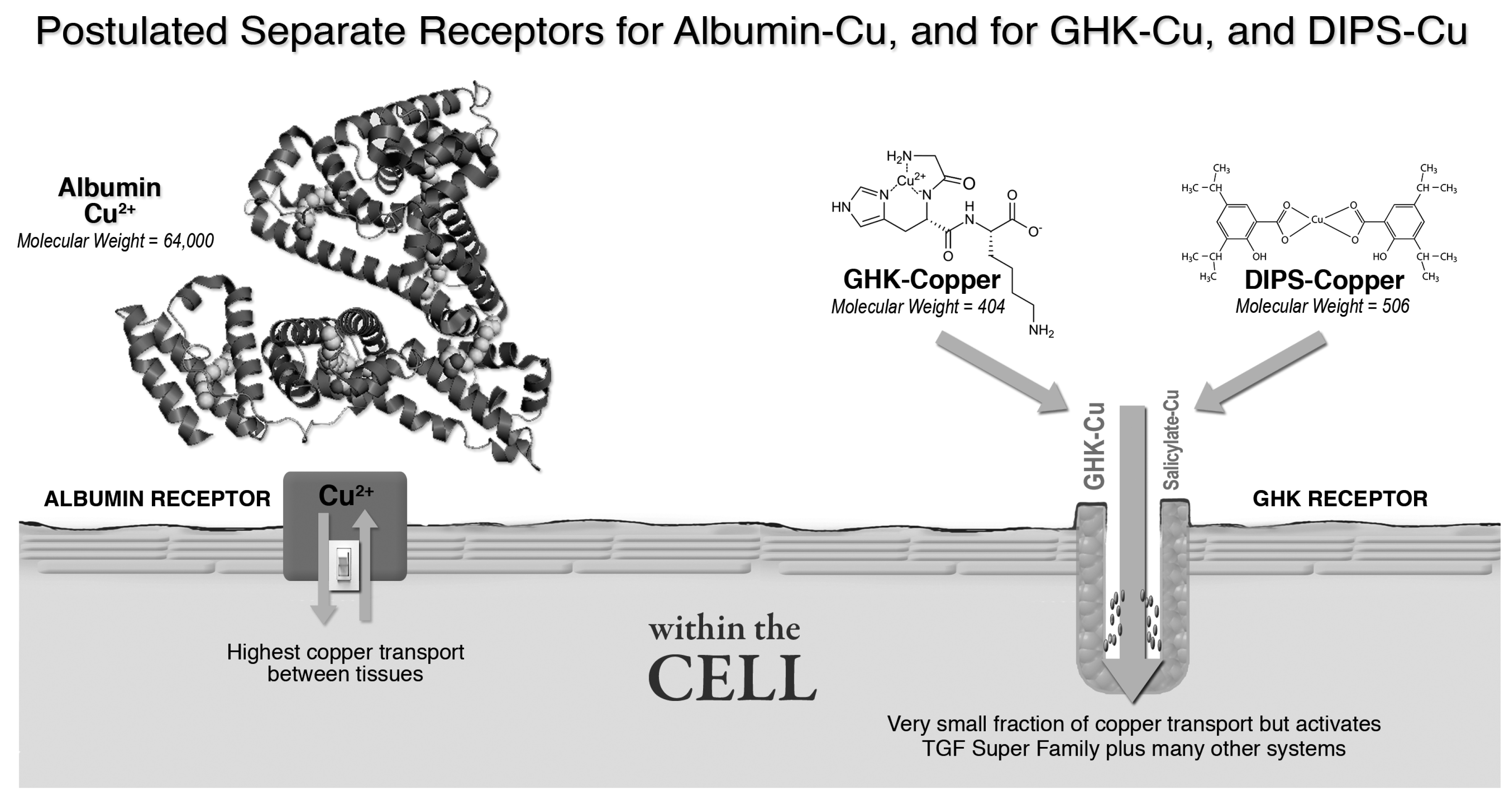
Product Image Context (How I Use It in Content)
If you’re building a page around this topic (mechanism-first, not claim-first), the best practice is to pair the image with a neutral explanation of what the peptide is being discussed in relation to. Here’s the provided product image:
How to Evaluate Future Evidence (A Short, Evidence-Driven Checklist)
If you’re reading new papers on GHK and gene expression, here’s a checklist I’ve used repeatedly to quickly separate “interesting” from “convincing.”
- Was gene expression directly measured? Prefer RNA-seq, qPCR panels, or credible transcriptomic profiling.
- Is there mechanistic pathway support? Look for pathway assays (e.g., transcription factor activity, signaling markers) that explain the transcriptional shift.
- Are results tied to nervous system function markers? Synaptic genes, neuroinflammation markers, mitochondrial stress signatures, or neurotrophic pathways are more relevant than generic stress-only signatures.
- Is there a clear discussion of limitations? Dose, cell type, copper context, and exposure duration should be addressed transparently.
- Do functional outcomes match the transcript changes? If gene expression moves but functional endpoints don’t, the mechanism story is weaker.
FAQ
Does GHK Cu peptide directly change genes in brain cells?
In many studies, the strongest support comes from experiments that directly measure gene expression after exposure in relevant cellular models. Whether the effect is “direct” versus mediated through signaling pathways varies by study design, so the most reliable evidence comes from mechanistic work that links exposure → pathway activity → transcriptional change in appropriate nervous system–relevant models.
How is “ghk cu peptide and cancer” related to nervous system decline?
The link is typically through shared biology such as oxidative stress balance, survival signaling, and inflammation—processes that also influence neuronal health. The limitation is context: what drives survival in one cell type or disease setting may not translate to neurodegeneration in a straightforward way, so cancer-adjacent findings should be interpreted as mechanistic hints rather than direct evidence for cognitive outcomes.
What would count as convincing evidence for cognitive decline relevance?
The most convincing evidence would connect gene expression changes to nervous system–relevant functional outcomes (e.g., reduced neuroinflammatory signatures, improved cell resilience, or synaptic maintenance indicators) in models that approximate the neurobiology of cognitive decline, ideally with consistent directionality and mechanistic pathway support.
Conclusion
The most useful way to think about GHK is as a regulator that can shift gene expression programs tied to oxidative stress responses, inflammation, survival pathways, and neurotrophic support—mechanisms that can matter for nervous system function and, by extension, cognitive decline. At the same time, the phrase ghk cu peptide and cancer points to shared cellular stress and survival biology, but it doesn’t automatically imply direct translation to brain outcomes; context, dose, and cell type are crucial.
Next step: Choose one recent paper that reports GHK-related gene expression changes, then apply the checklist above—direct transcription measurement, pathway logic, nervous system–relevant markers, and functional correlation—before drawing any mechanistic conclusions.
Discussion